Ultrasound In Scrotal Doppler Testicular And Scrotal Doppler Ultrasound
Ultrasound In Scrotal Doppler Testicular And Scrotal Doppler Ultrasound
If this picture is your intelectual property (copyright infringement) or child pornography / immature images, please Contact Us for abuse. We will follow up your report/abuse within 24 hours.
Related Images of figure 1 from the validity of grayscale and color doppler ultrasound in assessment of scrotal
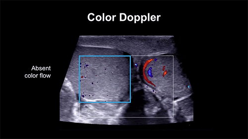
Figure 1 From The Validity Of Grayscale And Color Doppler Ultrasound In Assessment Of Scrotal
Figure 1 From The Validity Of Grayscale And Color Doppler Ultrasound In Assessment Of Scrotal

Figure 1 From The Validity Of Grayscale And Color Doppler Ultrasound In Assessment Of Scrotal
Figure 1 From The Validity Of Grayscale And Color Doppler Ultrasound In Assessment Of Scrotal
Figure 1 From The Validity Of Grayscale And Color Doppler Ultrasound In Assessment Of Scrotal
Figure 1 From The Validity Of Grayscale And Color Doppler Ultrasound In Assessment Of Scrotal
Figure 1 From The Validity Of Grayscale And Color Doppler Ultrasound In Assessment Of Scrotal
Figure 1 From The Validity Of Grayscale And Color Doppler Ultrasound In Assessment Of Scrotal

Table 1 From The Validity Of Grayscale And Color Doppler Ultrasound In Assessment Of Scrotal
Table 1 From The Validity Of Grayscale And Color Doppler Ultrasound In Assessment Of Scrotal
Figure 1 From Role Of Grayscale And Color Doppler Ultrasonography In Evaluation Of Scrotal
Figure 1 From Role Of Grayscale And Color Doppler Ultrasonography In Evaluation Of Scrotal
Figure 1 From Role Of Grayscale And Color Doppler Ultrasonography In Evaluation Of Scrotal
Figure 1 From Role Of Grayscale And Color Doppler Ultrasonography In Evaluation Of Scrotal

Table 3 From The Validity Of Grayscale And Color Doppler Ultrasound In Assessment Of Scrotal
Table 3 From The Validity Of Grayscale And Color Doppler Ultrasound In Assessment Of Scrotal

Figure 1 From Role Of Grayscale And Color Doppler Ultrasonography In Evaluation Of Scrotal
Figure 1 From Role Of Grayscale And Color Doppler Ultrasonography In Evaluation Of Scrotal

Table 3 From The Validity Of Grayscale And Color Doppler Ultrasound In Assessment Of Scrotal
Table 3 From The Validity Of Grayscale And Color Doppler Ultrasound In Assessment Of Scrotal

Pdf The Validity Of Grayscale And Color Doppler Ultrasound In Assessment Of Scrotal Swellings
Pdf The Validity Of Grayscale And Color Doppler Ultrasound In Assessment Of Scrotal Swellings

Scrotal Color Doppler Ultrasound Showing Subcutaneous Fat Layers Download Scientific Diagram
Scrotal Color Doppler Ultrasound Showing Subcutaneous Fat Layers Download Scientific Diagram

Cureus Evaluation Of Scrotal Pathologies By Ultrasound And Color Doppler
Cureus Evaluation Of Scrotal Pathologies By Ultrasound And Color Doppler
Testicularscrotal Doppler Protocol Sonographic Tendencies
Testicularscrotal Doppler Protocol Sonographic Tendencies

Testicularscrotal Doppler Protocol Sonographic Tendencies
Testicularscrotal Doppler Protocol Sonographic Tendencies

Control Scrotal Color Doppler Ultrasound A Resolution Of Venous Download Scientific Diagram
Control Scrotal Color Doppler Ultrasound A Resolution Of Venous Download Scientific Diagram
Endovascular Today Varicocele Embolization For Infertility April 2017
Endovascular Today Varicocele Embolization For Infertility April 2017
A Sagittal Grayscale Image Shows A Normal Testicle With Homogeneous Download Scientific Diagram
A Sagittal Grayscale Image Shows A Normal Testicle With Homogeneous Download Scientific Diagram

Testicularscrotal Doppler Protocol Sonographic Tendencies
Testicularscrotal Doppler Protocol Sonographic Tendencies

Ppt Scrotal Ultrasound Powerpoint Presentation Free Download Id5671926
Ppt Scrotal Ultrasound Powerpoint Presentation Free Download Id5671926
Testicularscrotal Doppler Protocol Sonographic Tendencies
Testicularscrotal Doppler Protocol Sonographic Tendencies

Grayscale A And Color Doppler B Ultrasound Images Showing An Download Scientific Diagram
Grayscale A And Color Doppler B Ultrasound Images Showing An Download Scientific Diagram

What Do The Colors Mean On Doppler Ultrasound Scan
What Do The Colors Mean On Doppler Ultrasound Scan
Figure 3 From Validity Of Ultrasound With Color Doppler To Differentiate Between Benign And
Figure 3 From Validity Of Ultrasound With Color Doppler To Differentiate Between Benign And

Grayscale A And C F And Colour Doppler Ultrasound B Show A Massive Download Scientific
Grayscale A And C F And Colour Doppler Ultrasound B Show A Massive Download Scientific

Grayscale Ultrasound With Color Doppler Demonstrates An Abnormal Color Download Scientific
Grayscale Ultrasound With Color Doppler Demonstrates An Abnormal Color Download Scientific

Neonatal Testicular Torsion A Transverse Grayscale Sonogram Of The Download Scientific
Neonatal Testicular Torsion A Transverse Grayscale Sonogram Of The Download Scientific

Figure 0001role Of Color Doppler In Scrotal Lesions Open I
Figure 0001role Of Color Doppler In Scrotal Lesions Open I
Best Predictors Of Grayscale Ultrasound Combined With Color Doppler In The Diagnosis Of Retained
Best Predictors Of Grayscale Ultrasound Combined With Color Doppler In The Diagnosis Of Retained
Figure 1 From Validity Of Ultrasound With Color Doppler To Differentiate Between Benign And
Figure 1 From Validity Of Ultrasound With Color Doppler To Differentiate Between Benign And
Figure 1 From Validity Of Ultrasound With Color Doppler To Differentiate Between Benign And
Figure 1 From Validity Of Ultrasound With Color Doppler To Differentiate Between Benign And
