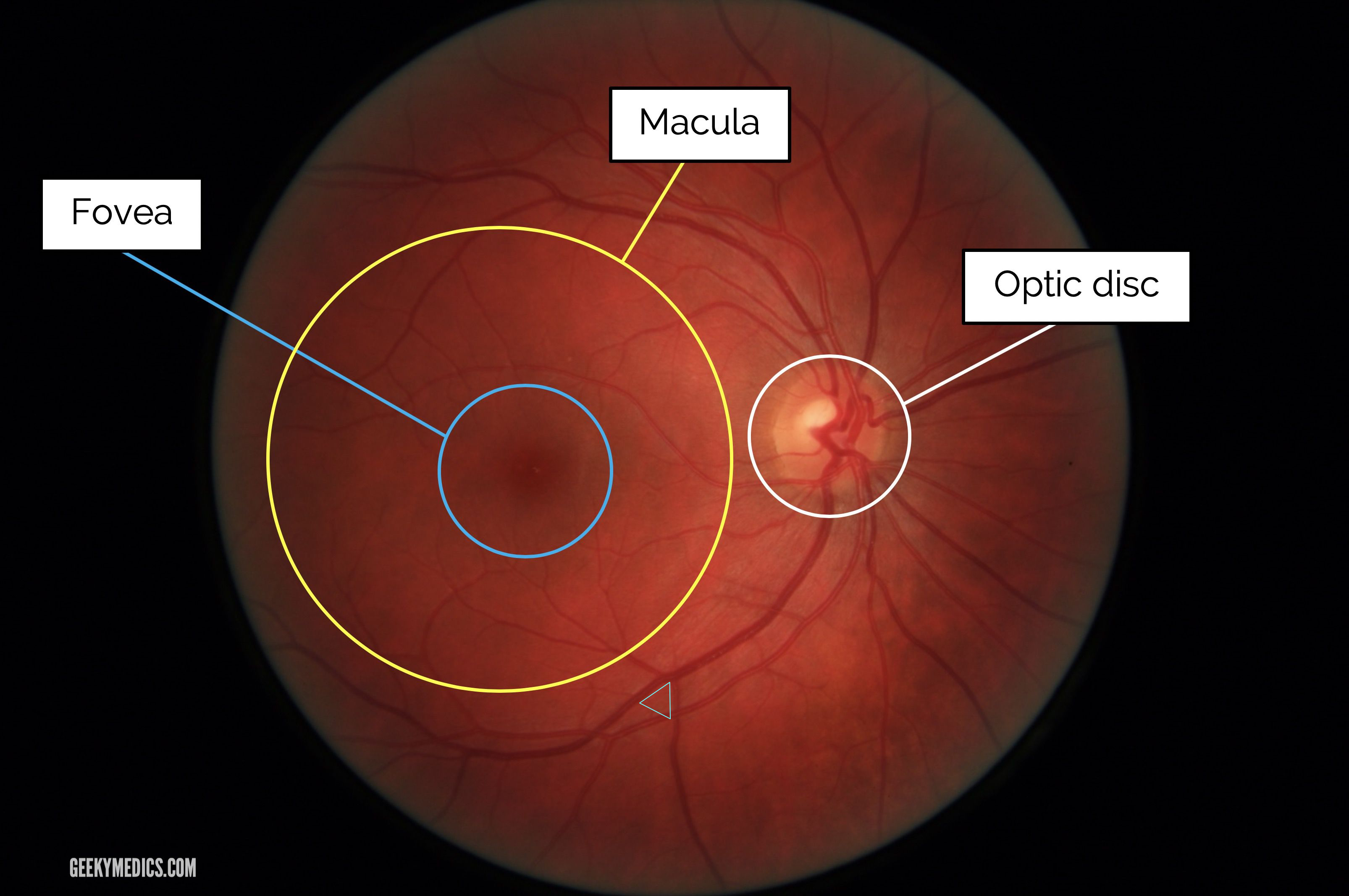
Fundoscopic Appearances Of Retinal Pathologies Geeky Medics
Fundoscopic Appearances Of Retinal Pathologies Geeky Medics
If this picture is your intelectual property (copyright infringement) or child pornography / immature images, please Contact Us for abuse. We will follow up your report/abuse within 24 hours.
Related Images of fundus photography of the right eye showing myelinated retinal nerve download scientific

Fundus Photography Of The Right Eye Showing Myelinated Retinal Nerve Download Scientific
Fundus Photography Of The Right Eye Showing Myelinated Retinal Nerve Download Scientific

Funduscopic Image Of The Right Eye Showing Myelinated Retinal Nerve Download Scientific Diagram
Funduscopic Image Of The Right Eye Showing Myelinated Retinal Nerve Download Scientific Diagram

Fundus Photography Of The Patients Right Eye Showed Large Cupped Download Scientific Diagram
Fundus Photography Of The Patients Right Eye Showed Large Cupped Download Scientific Diagram

Fundus Photography Of The Right Eye Showing Myelinated Retinal Nerve Download Scientific
Fundus Photography Of The Right Eye Showing Myelinated Retinal Nerve Download Scientific

Fundus Photography Showing A Normal Right Eye And Left Eye With Download Scientific Diagram
Fundus Photography Showing A Normal Right Eye And Left Eye With Download Scientific Diagram

Fundusphotographofnormalrighteye Doris Lu Optometrist
Fundusphotographofnormalrighteye Doris Lu Optometrist
Cureus Bilateral Peripapillary Retinal Nerve Fiber Layer Myelination In A 26 Year Old Man
Cureus Bilateral Peripapillary Retinal Nerve Fiber Layer Myelination In A 26 Year Old Man
Fundus Photography Of Right Eye Showing Optic Disc Edema And Retinal Download Scientific
Fundus Photography Of Right Eye Showing Optic Disc Edema And Retinal Download Scientific

Fundus Photography Of The Right Eye Taken About 2 Weeks After The Acute Download Scientific
Fundus Photography Of The Right Eye Taken About 2 Weeks After The Acute Download Scientific
A Fundus Photograph Of The Right Eye Showing A Dilated Retinal Vein Download Scientific
A Fundus Photograph Of The Right Eye Showing A Dilated Retinal Vein Download Scientific

Fundus Photographs Showing The Normal Appearance In The Right Eye And Download Scientific
Fundus Photographs Showing The Normal Appearance In The Right Eye And Download Scientific
Large Area Of Myelinated Retinal Nerve Fibers Ophthalmology
Large Area Of Myelinated Retinal Nerve Fibers Ophthalmology

Myelinated Nerve Fiber Layer Optic Nerve Retina Image Bank
Myelinated Nerve Fiber Layer Optic Nerve Retina Image Bank
Fundus Photos Of Both Eyes A Right Eye Showing Swollen And Hyperemic Download Scientific
Fundus Photos Of Both Eyes A Right Eye Showing Swollen And Hyperemic Download Scientific
Syndrome Of Myelinated Nerve Fibers Hyperopia Strabismus And Amblyopia Ophthalmology Retina
Syndrome Of Myelinated Nerve Fibers Hyperopia Strabismus And Amblyopia Ophthalmology Retina
Fundus Photographs Of Daughter Showing Peripapillary Myelinated Nerve Download Scientific
Fundus Photographs Of Daughter Showing Peripapillary Myelinated Nerve Download Scientific

Cureus Ocular Findings Associated With Myelinated Retinal Nerve Fibers
Cureus Ocular Findings Associated With Myelinated Retinal Nerve Fibers

Right Fundus Myelinated Nerve Fibers Temporal Optic Disc Pit And Download Scientific Diagram
Right Fundus Myelinated Nerve Fibers Temporal Optic Disc Pit And Download Scientific Diagram


Fundoscopic Appearances Of Retinal Pathologies Geeky Medics
Fundoscopic Appearances Of Retinal Pathologies Geeky Medics
Fundus Examination Of Both Eyes And Sd Oct Scan A Right Eye Re Download Scientific
Fundus Examination Of Both Eyes And Sd Oct Scan A Right Eye Re Download Scientific

The Spark Imagewise 28 Characteristics Of Myelinated Retinal Nerve Fiber Layer On Sdoct
The Spark Imagewise 28 Characteristics Of Myelinated Retinal Nerve Fiber Layer On Sdoct
Fundus Photo Montage Showing Normal Right And Left Eye Showing Download Scientific Diagram
Fundus Photo Montage Showing Normal Right And Left Eye Showing Download Scientific Diagram
A The Fundus Photograph Of Both Eyes Shows A Small Area Of Myelinated Download Scientific
A The Fundus Photograph Of Both Eyes Shows A Small Area Of Myelinated Download Scientific

Fundus Photograph Of The Right Eye Showing Branch Retinal Artery Download Scientific Diagram
Fundus Photograph Of The Right Eye Showing Branch Retinal Artery Download Scientific Diagram
Rare Association Of Optic Disk Pits With Myelinated Retinal Nerves Journal Of American
Rare Association Of Optic Disk Pits With Myelinated Retinal Nerves Journal Of American
